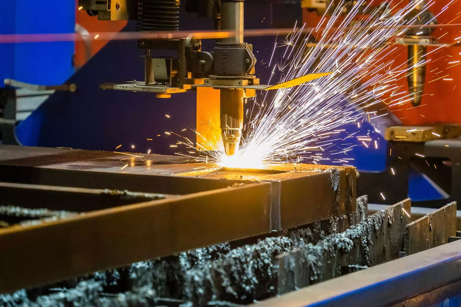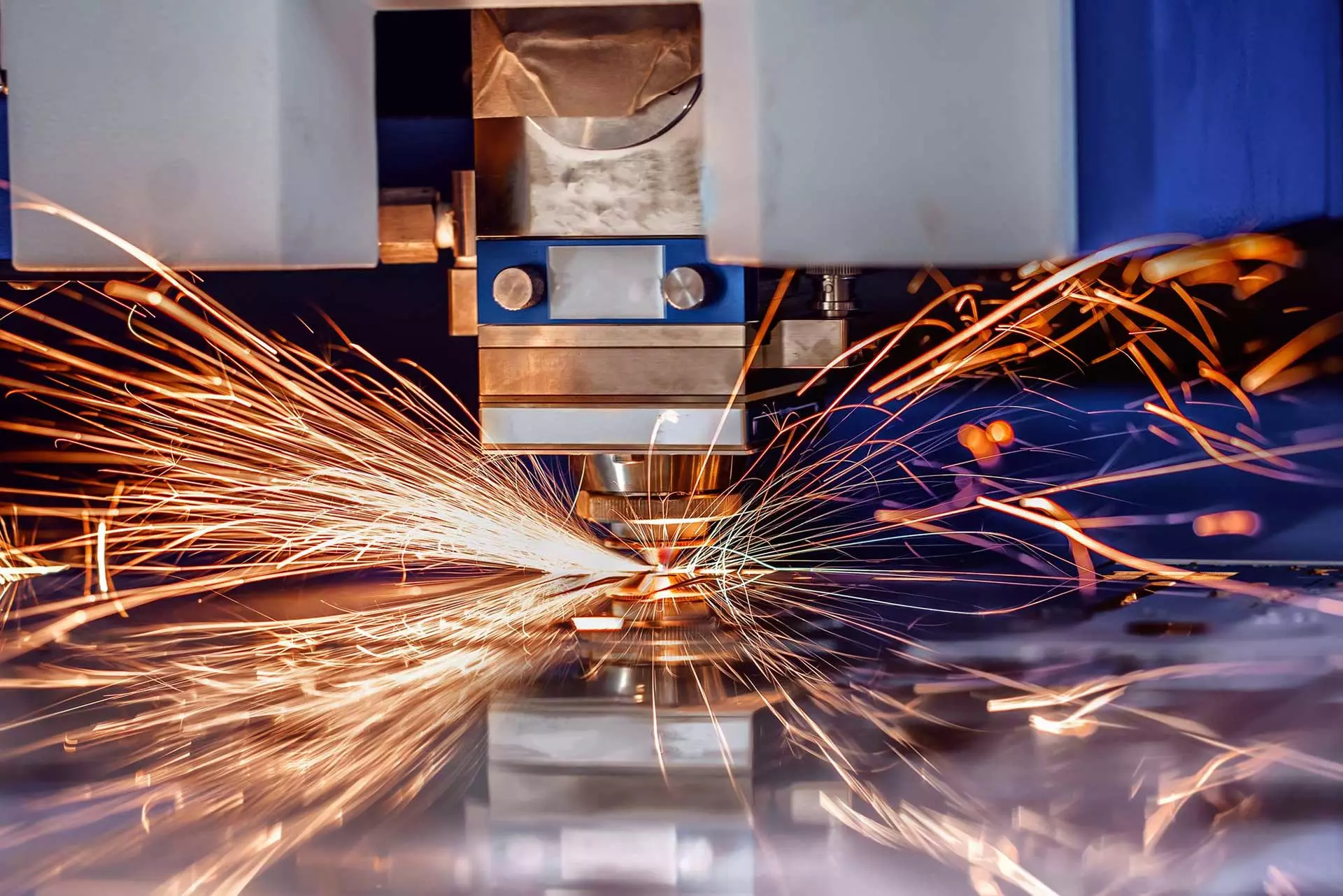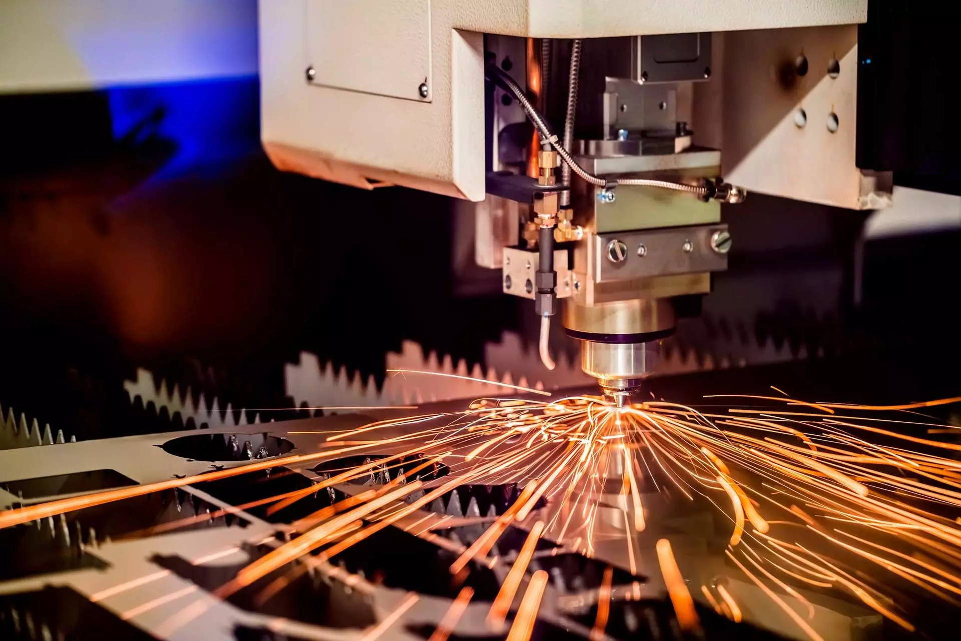


Digital Holographic Microscopy measures the form and displacement of objects by digitally reconstructing holograms captured during inspection of turbine components, making this technology ideal for inspection of turbine parts that have proven their ability to prevent compressor seal bond failures, providing manufacturers with significant cost savings.
DHM involves splitting a uniform laser beam into an object beam and reference beam that interact with a sample to scatter light in various directions, and recording this interference pattern using a light detector. Once recorded, this interference pattern is fed back into a computer which uses a reconstruction algorithm to calculate a viewable 3D image of it.
DHM involves splitting a uniform laser beam into an object beam and reference beam that interact with a sample to scatter light in various directions, and recording this interference pattern using a light detector. Once recorded, this interference pattern is fed back into a computer which uses a reconstruction algorithm to calculate a viewable 3D image of it.
Holography
Holography is an imaging technique that utilizes light to create three-dimensional (3D) images, often used in both art and science applications. Holography’s ability to capture spatial data at high resolution makes it an invaluable asset in scientific applications such as flow analysis and particle/cell dynamics and monitoring; additionally, this technique has great promise as medical diagnostics or drug discovery.
An image is created by shining a beam of light onto material which records diffraction patterns, which are then reconstructed to form the image. Laser light is often focused on a plate acting as a lens and its beam directed onto this plate for reconstruction of images on display devices, such as computer monitors. Once constructed, these holograms can also be scanned to produce higher-resolution images as well as stored digitally for future reference.
Holography technology does not work well during complex movements because the light must remain steady to achieve quality results. Furthermore, the process requires both expensive hardware and software for it to function successfully; additionally it takes many hours for 3D image production; equipment should not be moved during production process and must remain stationary on a flat surface that won’t vibrate when someone walks across or cars drive by outside; professional holography tables feature honeycomb-shaped support layers as well as pneumatic legs designed to dampen vibration.
Holography provides more than just spatial images; it also gives phase information about objects. This can help analyze object structures and their diffraction patterns, as well as estimate particle movement estimates. Holographic method applications range from early cell death research, detection of ionic regulation, to particle movement estimation estimations.
Moreover, this technology is more accurate than traditional medical imaging techniques. It can detect injuries to soft and hard tissues of patients more accurately while storing all previous images digitally, helping physicians easily analyze patient conditions more easily. Furthermore, its applications span cardiology, pulmonary, genitourinary, and musculoskeletal radiology as well as cancer detection in patients.
Reconstruction
Reconstruction is a computational technique used to reconstruct amplitude and phase images from measurements taken of objects. Reconstruction uses interference and diffraction as its basis; its key role in holography has yet to be fully appreciated due to the difficulty involved with performing calculations required; various schemes have been devised but none has achieved high lateral and depth resolution without using microscope objective lenses; these lenses tend to introduce significant vibrations which limit their usefulness for biological microscopy.
One of the key developments in digital holography is optical phase conjugation, which works to eliminate distortions caused by passing through an aberrating medium and can help free-space optical communications compensate for atmospheric turbulence, as well as being used for recording and displaying 3-D images.
Digital holography has many applications in medicine, and orthopaedics is particularly notable where doctors need to visualize bones and joints in real time for accurate diagnosis and more efficient patient treatment. Holography also can assist surgeons avoid errors during patient positioning to maximize surgery success rates.
Digital holograms contain all the information needed to reconstruct an object’s three-dimensional structure. As illustrated in Figure A, a laser illuminates a pinhole serving as a point source, producing spherical waves that interfere with reference waves recorded on photographic plate or film and produce the hologram image that contains both amplitude and phase information of an object’s three-dimensional form. Restoring such an hologram provides access to its information.
Digital holography is an exciting technology with potential medical applications, yet needs further refinement. For instance, digital holography can help monitor changes in cell density and permeability that would otherwise be difficult to track using traditional bright-field microscopes; and visualize movement of particles or specimens through fluid. Both features make digital holography beneficial in numerous areas including drug discovery and tissue engineering.
Image capturing system
Holography is a photographic technique that creates three-dimensional impressions of objects using radiation ranging from microwaves to x-rays. When illuminated, reflecting and transmitted waves combine into an interference pattern on photosensitive plates known as holograms; this interference pattern contains information about their shape and position that can be reproduced when illuminated with coherent light sources such as laser beams.
Dennis Gabor first developed this process in 1947 as a means to enhance electron microscopy resolution. Since then, his ideas have also applied to imaging with any coherent light source like lasers. Now we use digital holographic microscopy for recording and reconstructing images using computer processing; less costly than traditional optical microscopy techniques it allows us to examine a range of samples including proteins to cells.
Beating reflections with holography takes careful effort. A dark working space and vibration-free surroundings are required, as any such movements could blur interference fringes and distort your image. Furthermore, recording must be completed using an impressive laser. However, this task is no easy feat and often requires sophisticated laser systems and optical tables in order to achieve quality output – making holography previously an expensive endeavor; but Lloyd Cross was able to develop his own home-made solution that made holography both cheaper and more reliable than before.
One of the primary challenges associated with holograms is that their images don’t compare to traditional microscope pictures in terms of sharpness. A recent study demonstrated how deep learning-based upscaling can significantly enhance image quality of holograms by employing neural network algorithms to inspect their diffraction patterns and adjust reconstruction depth accordingly, offering another method for particle/cell dynamics analysis.
Image analysis
Image Analysis involves measuring the amplitude and phase of a hologram to produce an image, accomplished by recording its interference pattern on an image sensor, then analyzing it with computer software. This can assist users in identifying size and location of objects within samples as well as classification purposes. Furthermore, imaging techniques like this one allow researchers to observe cell dynamics without disrupting normal functioning of cells.
Lensless digital holographic microscopy is an exciting advancement in microscopy that holds much promise to replace traditional photochemical methods and open up a host of opportunities. However, this technology still faces certain hurdles to reaching its full potential; such as needing sufficient memory and processing power to calculate reconstructions accurately as well as being used alongside other microscopy techniques for optimal results.
Holography’s main advantage lies in its ability to detect and visualize objects with varied optical properties, like living cells. Holography technology is particularly useful for viewing transparent objects, such as living cells. By measuring optical thickness variations between objects, transmission holography provides excellent analysis capabilities while reflection holography allows one to analyze defects within transparent samples.
Holographic imaging differs from other microscopy techniques by being capable of producing two images: an amplitude image and phase image. The former represents object brightness while its optical thickness can be determined from this phase image; hence why this technique is often known as quantitative phase contrast microscopy (QPCM). QPCM can also be used to examine unstained cells to understand their structure and functions better.
Recent research demonstrated how the combination of holographic microscopy and deep learning can greatly enhance image reconstruction quality and classification accuracy, as well as reduce reconstruction times and storage needs significantly. According to its authors, such advances could prove highly advantageous in life sciences and medical fields.